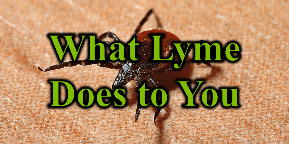
Welcome to the seventh essay in our special series sharing insights and recent research from the MEDMAPS 2023 Spring Conference attended by Dr. Potter. MAPS stands for Medical Academy for Pediatric Special Needs and arose from the original Defeat Autism Now organization initially serving parents and providers caring for children with autism spectrum disorder). This Spring Conference focused not only on autism research but on Lyme and other infections including COVID’s effects on children. Come back in the coming weeks to read more about what I learned at the conference so that Sanctuary can provide cutting edge care to your precious little ones.
Our patients with Lyme disease usually want to really understand their illness. At Sanctuary and within the world of functional medicine, we believe that all should have a basic understanding of what is making them ill, what is stealing their optimal life from them. Knowing the enemy helps us know how to fight. Understanding the mechanisms through which Lyme causes a variety of bodily dysfunctions will equip you to fight back. With Lyme this will take some work as its attack mechanisms are quite complicated. For those of you who are facing Lyme disease, this understanding will also encourage you that you can recover by addressing the variety of mechanisms used by Lyme to make you feel miserable.
While several pathways participate in the pathogenesis of Lyme symptoms, at the bottom of all the symptoms and spread lies inflammation. Once the spirochete bacteria behind Lyme enters the body through the bite of a tick or other means, various substances begin building inflammation which then allows the bacteria to spread, grow, and invade further. Borrelia, the bacteria behind Lyme, are attracted to connective tissues in our bodies as they are made of collagen, aggrecan, and hyaluronic acid (which borrelia uses for food). Inflammation triggered by the borrelia release enzymes and trigger processes which breakdown these tissues so Borrelia can feed easier.
Lyme Borrelia begin their invasion with the help of tick saliva and its various immune altering components. Some of the chemicals inhibit interleukin 8 (Hajnicka, 2001), while some inhibit interleukin 12 (Anguita, 1996), and some inhibit Interferon gamma (Dame,2007). Ultimately they shift our immune system from a Th1 cellular response to a Th2 antibody response thus inhibiting nitric oxide production. With longer feeding times of the tick in the skin, more saliva means more immune shifting allowing more borrelia invasion.
Once the Lyme Borrelia enters the body with the help of the tick and its saliva, further inflammatory processes ensue. On the flagella of the bacteria, a protein called flagellin stimulate NF-kB (nuclear factor kappa beta) which then triggers further inflammatory processes. Immune cells proliferate and increase the break-down of tissues in order to allow the cells to reach the infection. This also opens pathways for the Borrelia to spread along the same lines of disrupted connective tissue and feed on the broken-down tissues.
The participating inflammation pathways include MAPK’s (mitogen activated protein kinases), ERKs (types of protein-serine/threonine kinases), JNKs (c-Jun N-terminal kinases) (Johnson, 2002), and p38 kinases (Johnson, 2022). They include IDO (Indoleamine 2,3 dioxygenase (Love, 2015) which starts a pathway leading to decreased T cells and impairs brain function by decreasing melatonin and serotonin. Interleukin 8 is triggered and leads to inflammation and cellular damage. Interleukin 1B raises, stimulating cell proliferation (Molina-Holgado, 2000) and increases sensitivity to pain (Simon, 1999). Along with this, interleukin 6 rises, crosses the Blood Brain Barrier (BBB) and alters the body’s temperature regulation system (Egecioglu, 2018) besides affecting hormonal balance (Spath-Schwalbe, 1994), and leading to neuronal degeneration (Kimura, 2010). If that were not enough, Tumor Necrosis Factor alpha rises, further imbalancing hormones (Dunn, 2000), affecting metabolic systems (Knobler, 2005) and our nervous system (Raffaele, 2020). One more pathway are the matrix metalloproteinases which break down collagen not only allowing Borrelia to feed, but also causing a lot of damage to us.
These pathways enable the initial infection to take root and if these pathways are not arrested, eventually set the body up for chronic infections. By hijacking the immune system, Borrelia sabotage the defenses and evade the counterattacks. When conventional medicine focuses solely on attacking the invaders, the Borrelia have an easy time by using our bodies’ mechanisms against the conventional approaches. Only when our immune systems are engaged and optimized can the infection be fully overcome (Bernard, 2018).
We can employ various natural remedies to modulate these sabotaged pathways. First, we must remove other contributors to inflammation in our body like inflammatory food choices and toxins. Second, we must modify lifestyle in terms of stress and sleep to prepare our bodies for proper responses.
Then we can utilize the natural therapies God has provided. TNF alpha can be lowered by herbs such as Cordyceps (Zhu, 2012) and Houttuynia (Park, 2005). P38 and MAPK can also be modified by Cordyceps (Das, 2021) or Japanese knotweed (Kim, 2013). IL-1B is also lowered by Japanese knotweed and Cordyceps. IL-6 is lowered by Skullcap (Liu, 2019) and other herbals. Skullcap also inhibits IDO. Beyond that modified citrus pectin can decrease the Galectin 3 pathway which perpetuates inflammation.
Combining natural and pharmaceutical options increases the success of Lyme therapy. Given the complexity and breadth of Borrelia’s mechanisms of invasion, it is not surprising that multiple responses are needed to successfully eradicate Lyme. Dr. Hinchey’s talk went much deeper at the conference and is a worthwhile investment if you want to attend her upcoming Lyme conference found on LymeBytes website. Meanwhile, if you are suffering with Lyme and just want someone who will not hold back in guiding you through the maze of these mechanisms impacting your daily life, Sanctuary is here for you, applying the best of what we have available and constantly seeking to advance the efficacy of our methods with these and other conferences. Helping our patients live healthier, more abundant lives requires this diligence towards excellence.
Medical Disclaimer: These essays are for educational purposes only. We assume no responsibility for your choice to implement something from these essays. Even if you are a patient of our clinic, you should consult with us before adding therapies. If you are not one of our patients, talk with your health care provider before trying any of these therapies.
Bibliography:
Hajnická, V., Kocáková, P., Sláviková, M., Slovák, M., Gasperík, J., Fuchsberger, N., & Nuttall, P. A. (2001). Anti-interleukin-8 activity of tick salivary gland extracts. Parasite immunology, 23(9), 483–489. https://doi.org/10.1046/j.1365-3024.2001.00403.x
Anguita, J., Persing, D. H., Rincon, M., Barthold, S. W., & Fikrig, E. (1996). Effect of anti-interleukin 12 treatment on murine lyme borreliosis. The Journal of clinical investigation, 97(4), 1028–1034. https://doi.org/10.1172/JCI118494
Dame, T. M., Orenzoff, B. L., Palmer, L. E., & Furie, M. B. (2007). IFN-gamma alters the response of Borrelia burgdorferi-activated endothelium to favor chronic inflammation. Journal of immunology (Baltimore, Md. : 1950), 178(2), 1172–1179. https://doi.org/10.4049/jimmunol.178.2.1172
Johnson, G. L., & Lapadat, R. (2002). Mitogen-activated protein kinase pathways mediated by ERK, JNK, and p38 protein kinases. Science (New York, N.Y.), 298(5600), 1911–1912. https://doi.org/10.1126/science.1072682
Russell, T. M., & Johnson, B. J. (2013). Lyme disease spirochaetes possess an aggrecan-binding protease with aggrecanase activity. Molecular microbiology, 90(2), 228–240. https://doi.org/10.1111/mmi.12276
Love, A. C., Schwartz, I., & Petzke, M. M. (2015). Induction of indoleamine 2,3-dioxygenase by Borrelia burgdorferi in human immune cells correlates with pathogenic potential. Journal of leukocyte biology, 97(2), 379–390. https://doi.org/10.1189/jlb.4A0714-339R
Molina-Holgado, E., Ortiz, S., Molina-Holgado, F., & Guaza, C. (2000). Induction of COX-2 and PGE(2) biosynthesis by IL-1beta is mediated by PKC and mitogen-activated protein kinases in murine astrocytes. British journal of pharmacology, 131(1), 152–159. https://doi.org/10.1038/sj.bjp.0703557
imon L. S. (1999). Role and regulation of cyclooxygenase-2 during inflammation. The American journal of medicine, 106(5B), 37S–42S. https://doi.org/10.1016/s0002-9343(99)00115
Egecioglu, E., Anesten, F., Schéle, E., & Palsdottir, V. (2018). Interleukin-6 is important for regulation of core body temperature during long-term cold exposure in mice. Biomedical reports, 9(3), 206–212. https://doi.org/10.3892/br.2018.1118
Kimura, A., & Kishimoto, T. (2010). IL-6: regulator of Treg/Th17 balance. European journal of immunology, 40(7), 1830–1835. https://doi.org/10.1002/eji.201040391
Dunn A. J. (2000). Cytokine activation of the HPA axis. Annals of the New York Academy of Sciences, 917, 608–617. https://doi.org/10.1111/j.1749-6632.2000.tb05426.x
Knobler, H., & Schattner, A. (2005). TNF-{alpha}, chronic hepatitis C and diabetes: a novel triad. QJM : monthly journal of the Association of Physicians, 98(1), 1–6. https://doi.org/10.1093/qjmed/hci001
Raffaele, S., Lombardi, M., Verderio, C., & Fumagalli, M. (2020). TNF Production and Release from Microglia via Extracellular Vesicles: Impact on Brain Functions. Cells, 9(10), 2145. https://doi.org/10.3390/cells9102145
Quentin Bernard, Alexis A. Smith, Xiuli Yang, Juraj Koci, Shelby D. Foor, Sarah D. Cramer, Xuran Zhuang, Jennifer E. Dwyer, Yi-Pin Lin, Emmanuel F. Mongodin, Adriana Marques, John M. Leong, Juan Anguita, Utpal Pal. Plasticity in early immune evasion strategies of a bacterial pathogen. Proceedings of the National Academy of Sciences, 2018; 201718595 DOI: 10.1073/pnas.1718595115
Zhu, Z. Y., Chen, J., Si, C. L., Liu, N., Lian, H. Y., Ding, L. N., Liu, Y., & Zhang, Y. M. (2012). Immunomodulatory effect of polysaccharides from submerged cultured Cordyceps gunnii. Pharmaceutical biology, 50(9), 1103–1110. https://doi.org/10.3109/13880209.2012.658114
Park, S. Y., Jin, M. L., Kang, N. J., Park, G., & Choi, Y. W. (2017). Anti-inflammatory effects of novel polygonum multiflorum compound via inhibiting NF-κB/MAPK and upregulating the Nrf2 pathways in LPS-stimulated microglia. Neuroscience letters, 651, 43–51. https://doi.org/10.1016/j.neulet.2017.04.057
Das, G., Shin, H. S., Leyva-Gómez, G., Prado-Audelo, M., Cortes, H., Singh, Y. D., Panda, M. K., Mishra, A. P., Nigam, M., Saklani, S., Chaturi, P. K., Martorell, M., Cruz-Martins, N., Sharma, V., Garg, N., Sharma, R., & Patra, J. K. (2021). Cordyceps spp.: A Review on Its Immune-Stimulatory and Other Biological Potentials. Frontiers in pharmacology, 11, 602364. https://doi.org/10.3389/fphar.2020.602364
Kim, J. H., Bae, C. H., Park, S. Y., Lee, S. J., & Kim, Y. (2010). Uncaria rhynchophylla inhibits the production of nitric oxide and interleukin-1β through blocking nuclear factor κB, Akt, and mitogen-activated protein kinase activation in macrophages. Journal of medicinal food, 13(5), 1133–1140. https://doi.org/10.1089/jmf.2010.1128
Liu, L., Dong, Y., Shan, X., Li, L., Xia, B., & Wang, H. (2019). Anti-Depressive Effectiveness of Baicalin In Vitro and In Vivo. Molecules (Basel, Switzerland), 24(2), 326. https://doi.org/10.3390/molecules24020326
Sanctuary Functional Medicine, under the direction of Dr Eric Potter, IFMCP MD, provides functional medicine services to Nashville, Middle Tennessee and beyond. We frequently treat patients from Kentucky, Alabama, Mississippi, Georgia, Ohio, Indiana, and more... offering the hope of healthier more abundant lives to those with chronic illness.


